Home
/ Labeled Plant Cell Under Electron Microscope / Plant Bodies Cells / A typical animal cell (as seen in an electron microscope) medical images for powerpoint 1.
Labeled Plant Cell Under Electron Microscope / Plant Bodies Cells / A typical animal cell (as seen in an electron microscope) medical images for powerpoint 1.
Labeled Plant Cell Under Electron Microscope / Plant Bodies Cells / A typical animal cell (as seen in an electron microscope) medical images for powerpoint 1.. All information about labeled plant cell under electron microscope. Typical animal cell pinocytotic vesicle lysosome golgi vesicles golgi vesicles rough er (endoplasmic reticulum) smooth er (no ribosomes) cell (plasma) membrane mitochondrion golgi apparatus nucleolus nucleus centrioles (2) each composed of 9 microtubule triplets microtubules cytoplasm ribosome Best way to observe blood under the microscope. A cell is a very tiny structure which exists in living bodies. Endoplasmic reticulum rough and smooth british society for.
Here's a diagram of a plant cell: Here's a diagram of a plant cell: Some of these differences can be clearly understood when the cells are examined under an electron microscope. Labeled animal cell under electron microscope. Cell structure teaching resources the science teacher, organelles biology for majors i, 11 different types of cells in the human body, class test, chronic inflammation under the microscope learn share.
Eukaryotic Cells Biology I from s3-us-west-2.amazonaws.com Microscope images in this course come from the light microscope (magnification up to 400x) and the electron microscope (magnification up to 500000x). Under the microscope, plant cells are seen as large rectangular interlocking blocks. Two main regions can be recognized in a. Electron microscopes use a beam of electrons instead of beams or rays of light. Organelle location, size, shape and position Therefore, we must use a microscope to visualize cells in a tissue. Typical animal cell pinocytotic vesicle lysosome golgi vesicles golgi vesicles rough er (endoplasmic reticulum) smooth er (no ribosomes) cell (plasma) membrane mitochondrion golgi apparatus nucleolus nucleus centrioles (2) each composed of 9 microtubule triplets microtubules cytoplasm ribosome (ii) presence of large central vacuole in plant cell.
Are plant cells prokaryotic or eukaryotic?
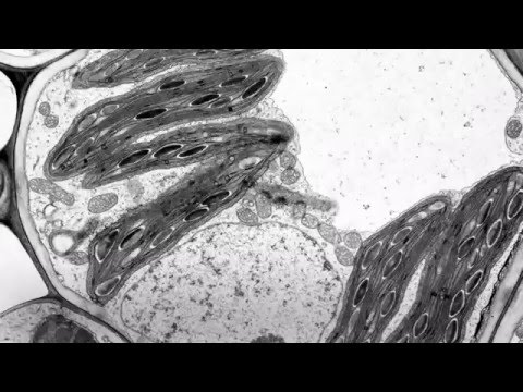
Electron microscopes use a beam of electrons instead of beams or rays of light. Typical animal cell pinocytotic vesicle lysosome golgi vesicles golgi vesicles rough er (endoplasmic reticulum) smooth er (no ribosomes) cell (plasma) membrane mitochondrion golgi apparatus nucleolus nucleus centrioles (2) each composed of 9 microtubule triplets microtubules cytoplasm ribosome In truth, there are still features of plant and anim. (1) they may be spheroidal, ovoid, stellate or collar shaped and differ in size and number in different cells. A plant cell as seen under electron microscope major differences between a plant cell and on animal cell are (i) presence of chloroplast in plant cell. For example, something that you draw as 3cm long, may in fact be 10, 000 times smaller in real life. The cell membrane, also known as plasma membrane or plasmalemma consists of three layers when viewed under the electron microscope. When viewing onion cells under a microscope a few drops of a certain solution are added to stain the cells and show these cells more clearly. All information about labeled plant cell under electron microscope. Here's a photo of a plant cell under an electron microscope. Photosynthetic cells of the leaf of elodea. Plant cell under a microscope labeled labelled diagram of a plant cell under a microscope. Plant cell under the microscope.
With the unaided eye, one can only see exceptionally large cells, such as the human ovum, which has a diameter of 100 µm. Pictures of labeled plant cell under electron microscope and many more. The electron micrograph of plastids: Best way to observe blood under the microscope. Plant cell biology structure based on their tissue:
1 2 Skill Interpretation Of Electron Micrographs Youtube from i.ytimg.com Labeled animal cell under electron microscope. Cell study with a light microscope. Coloured scanning electron micrograph (sem) of a blood vessel (arteriole) in the dermis of the skin. When viewing onion cells under a microscope a few drops of a certain solution are added to stain the cells and show these cells more clearly. Announcements results day is nearly here! The cell organelles are seen as tiny dots throughout the cytoplasm, whereas the nucleus is seen as a thick drop. Its this cell membrane that will contain all of the different parts of the cell. The cell is the basic structural and functional unit of life in all living organisms.
Cell structure teaching resources the science teacher, organelles biology for majors i, 11 different types of cells in the human body, class test, chronic inflammation under the microscope learn share.
Plant cell under a microscope labeled labelled diagram of a plant cell under a microscope. When viewing onion cells under a microscope a few drops of a certain solution are added to stain the cells and show these cells more clearly. Plant cell electron microscope google images teaching education plants life biology plant. These cell organelles perform specific functions within the cell. A typical animal cell (as seen in an electron microscope) medical images for powerpoint 1. The structural and functional elements of a plant cell have been revealed. Observe the labeled diagram of plant cell structure as given below. Make your work easier by using a label. Cell structure teaching resources the science teacher, organelles biology for majors i, 11 different types of cells in the human body, class test, chronic inflammation under the microscope learn share. Plant cell under the microscope. When viewing onion cells under a microscope a few drops of a certain solution are added to stain the cells and show these cells more clearly. Asked nov 28, 2017 in class. See how a generalized structure of an animal cell and plant cell look with labeled diagrams.
All information about labeled plant cell under electron microscope. Labels are a means of identifying a product or container through a piece of fabric, paper, metal or plastic film onto which information about them is printed. Bookfanatic89 diagram of plant cell under electron microscope. Draw and label an animal cell as seen under an electron microscope. Coloured scanning electron micrograph (sem) of a blood vessel (arteriole) in the dermis of the skin.
Gce Cie Biology Animal And Plant Cell Structures And Functions Biology Gce Cie Organelles Cell Function Eukaryotes from user-images.strikinglycdn.com The electron micrograph of plastids: Two main regions can be recognized in a. The diagram is very clear, and labeled; With the unaided eye, one can only see exceptionally large cells, such as the human ovum, which has a diameter of 100 µm. A plant cell as seen under electron microscope major differences between a plant cell and on animal cell are (i) presence of chloroplast in plant cell. Coloured scanning electron micrograph (sem) of a blood vessel (arteriole) in the dermis of the skin. Labelled diagram of plant cell under electron microscope (em image)? Its this cell membrane that will contain all of the different parts of the cell.
It uses a beam of electrons to illuminate the specimen instead of light as in the case of light microscope.
Labeled animal cell under electron microscope. Labelled diagram of plant cell under electron microscope (em image)? The electron microscope is more powerful than the light microscope. Best way to observe blood under the microscope. Here's a photo of a plant cell under an electron microscope. But at the same time it is interpretive. See how a generalized structure of an animal cell and plant cell look with labeled diagrams. Endoplasmic reticulum rough and smooth british society for. The organelles in a plant cell vary in size. However, no obvious structural damage was apparent, and several repeated scans gave the same images. See how a generalized structure of an animal cell and plant cell look with labeled diagrams. A cell is a very tiny structure which exists in living bodies. Cells and their surrounding cell membrane are usually very small because.
Share :
Post a Comment
for "Labeled Plant Cell Under Electron Microscope / Plant Bodies Cells / A typical animal cell (as seen in an electron microscope) medical images for powerpoint 1."


Post a Comment for "Labeled Plant Cell Under Electron Microscope / Plant Bodies Cells / A typical animal cell (as seen in an electron microscope) medical images for powerpoint 1."